Using advanced imaging techniques, scientists can peer inside chemical systems to explore their structure and behavior at the microscopic level. Such research can provide insights that aid in the search for promising new materials for important applications, ranging from batteries to semiconductors.
A project underway at the U.S. Department of Energy’s (DOE) Argonne National Laboratory seeks nothing less than to demonstrate the theoretical limits of the visualization of matter—that is, to identify the smallest features in objects that can be imaged and to what degree of detail.
Leveraging resources from the Argonne Leadership Computing Facility (ALCF) and Advanced Photon Source (APS), and led by researchers from the University of Michigan, the project aims to overcome experimental limitations and improve image quality in the characterization of materials. To this end, the team is building on algorithmic advances that enable heightened visual resolution, thereby improving scientists’ ability to identify and explore important chemical information. The ALCF and APS are U.S. DOE Office of Science user facilities.
Given the prominent role of materials science in disciplines spanning engineering, chemistry and physics, such advances stand to impact a wide range of technologies, including clean-energy essentials, like photovoltaics.
Algorithmic image reconstruction
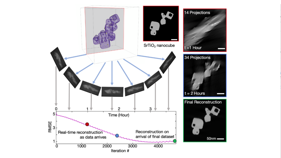
Electron and x-ray tomography are techniques for obtaining three-dimensional structures of specimens based on 2D projection images from electron and x-ray microscopy. Advances in tomographic image reconstruction stand to generate images of tiny matter that weren’t previously possible. In a paper recently published in npj Computational Materials, the researchers establish a limit-pushing method for creating high-resolution visualizations of atomic structure. The technique fills in visual gaps with information inferred by bombarding the targeted matter with electrons, producing differences in energy absorption that can serve as a sort of chemical fingerprint.
More specifically, the paper introduces multimodal electron microscopy, a technique for achieving high signal-to-noise ratio (SNR) recovery of material chemistry at nano- and atomic-scale resolutions. SNR measures the fidelity of a signal, with a higher ratio providing scientists with a clearer picture of the system.
“Our multimodal approach combines signals from an array of detectors to produce more detailed images than were previously possible,” said Robert Hovden, a professor at the University of Michigan and principal investigator (PI) of the project supported by the ALCF Data Science Program.
“The composite of data that’s harvested can help us recover, with a really high signal-to-noise ratio, the chemistry of whatever system we’re examining. Learning the ratios of the elements involved used to be quite challenging, but we can now identify a system’s individual atoms and assign each one a chemical composition. Not only that, we can obtain the amount of chemical composition—the stoichiometry, that is.” Stoichiometry specifically refers to the quantities of reactant and product substances in a chemical compound reaction.
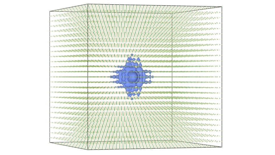
Indeed, the method for SNR recovery was tested against experimental data and computational simulations run on the ALCF’s Theta supercomputer. Stoichiometry can now be determined by leveraging data gleaned by scattering electrons off a target material.
Hovden’s team worked closely with Argonne staff to develop and test the computational viability of the project’s governing algorithm.
The simulations represent one of two computationally expensive components integral to the research, the other being multimodal reconstruction.
“Extremely large simulations were required. The images generated by these simulations are deceptively simple-looking, when in fact every pixel is assembled from thousands of quantum-mechanical calculations,” Hovden said.
More efficient signal interpretation
The work developed out of a desire to implement scattering physics techniques across different length scales. In physics, scattering refers to changes in the trajectory of a particle due to a collision with another particle.
“The x-ray community had been doing this kind of work, and given that using electron beams produces scattering physics roughly analogous to that of x-ray length-scales, it seemed likely some of those approaches might apply to the research I was doing,” Hovden said.
Current tomographic methods for interpreting atomic structure from elastic scattering signals fail to adequately describe the chemistry of a given specimen.
Similarly, chemical composition is determined using scattering signals derived from electron energy loss and/or energy-dispersive x-ray interactions, but current techniques tend to require electron doses so large that they produce noise or even damage the sample they’re being used to examine.
“Multimodal electron microscopy, by comparison, substantially improves SNR for chemical maps, with gains in the range of 300 to 500 percent frequently demonstrated,” said Huihuo Zheng, a computer scientist at the ALCF and co-PI on the project. “Moreover, the method can reduce electron doses by more than an order of magnitude, which translates to reduced environmental perturbation.”
Metrics obtained from the simulations help predict when and to what extent the approach fails.
Beyond its step forward in combining detectors and computational techniques to reconstruct particles, impact from the work is evident at APS.
“Put simply, achieving the higher resolutions we want to attain will enable the imaging of materials that we currently cannot image,” said co-PI Yi Jiang, an Argonne beamline data scientist working at the APS and a co-author of the paper. “We’ve already begun adapting similar multimodal image processing techniques for x-ray microscopy.”
Moving forward, Hovden and his team intend to extend their method to three dimensions.
“This really involves two questions,” Hovden said. “First there’s the matter of whether this dramatic improvement in SNR that enables visualization of atomic structure can also enable 3D visualization of full chemistry. If the answer is yes, then we have to discover at what resolution and for which classes of materials it’s true.”
==========
The Argonne Leadership Computing Facility provides supercomputing capabilities to the scientific and engineering community to advance fundamental discovery and understanding in a broad range of disciplines. Supported by the U.S. Department of Energy’s (DOE’s) Office of Science, Advanced Scientific Computing Research (ASCR) program, the ALCF is one of two DOE Leadership Computing Facilities in the nation dedicated to open science.
Argonne National Laboratory seeks solutions to pressing national problems in science and technology. The nation's first national laboratory, Argonne conducts leading-edge basic and applied scientific research in virtually every scientific discipline. Argonne researchers work closely with researchers from hundreds of companies, universities, and federal, state and municipal agencies to help them solve their specific problems, advance America's scientific leadership and prepare the nation for a better future. With employees from more than 60 nations, Argonne is managed by UChicago Argonne, LLC for the U.S. Department of Energy's Office of Science.
The U.S. Department of Energy's Office of Science is the single largest supporter of basic research in the physical sciences in the United States and is working to address some of the most pressing challenges of our time. For more information, visit https://energy.gov/science